Amritpal SinghDr. Amritpal Singh is a physician-scientist working at the intersection of medicine, computer science, and artificial intelligence. He holds an M.B.B.S. from Maulana Azad Medical College and an MS in Computer Science from Georgia Tech, where he specialized in machine learning, deep learning, reinforcement learning, graph methods, and medical robotics. His research bridges clinical medicine and AI, focusing on multimodal biomarker discovery, predictive modeling, and decision-support systems using imaging, genomics, and other patient data. Dr. Amritpal Singh is a physician-scientist working at the intersection of medicine, computer science, and artificial intelligence. He holds an M.B.B.S. from Maulana Azad Medical College and an MS in Computer Science from Georgia Tech, where he specialized in machine learning, deep learning, reinforcement learning, graph methods, and medical robotics. His research bridges clinical medicine and AI, focusing on multimodal biomarker discovery, predictive modeling, and decision-support systems using imaging, genomics, and other patient data. He is currently pursuing a PhD in Computer Science and Informatics at Emory University under Dr. Anant Madabhushi. His work develops clinically deployable AI systems that integrate radiology, digital pathology, genomics, and ocular imaging to improve risk prediction, early detection, and outcome modeling in cancer, cardiovascular disease, and other complex conditions. His broader interests include explainable AI, continual learning for healthcare, and building models that generalize across populations and institutions. He is currently pursuing a PhD in Computer Science and Informatics at Emory University under Dr. Anant Madabhushi. His work develops clinically deployable AI systems that integrate radiology, digital pathology, genomics, and ocular imaging to improve risk prediction, early detection, and outcome modeling in cancer, cardiovascular disease, and other complex conditions. His broader interests include explainable AI, continual learning for healthcare, and building models that generalize across populations and institutions. Dr. Singh has contributed to first-author publications at NeurIPS, MICCAI, and major clinical conferences, and has led projects translating AI research into clinically actionable tools. Dr. Singh has contributed to first-author publications at NeurIPS, MICCAI, and major clinical conferences, and has led projects translating AI research into clinically actionable tools. Email / GitHub / Google Scholar / LinkedIn / ORCID / Twitter / Kaggle |

|
News
|
ResearchI'm interested in artificial intelligence, machine learning for healthcare, computational pathology, radiology. Below are some of my recent research publications. |
|
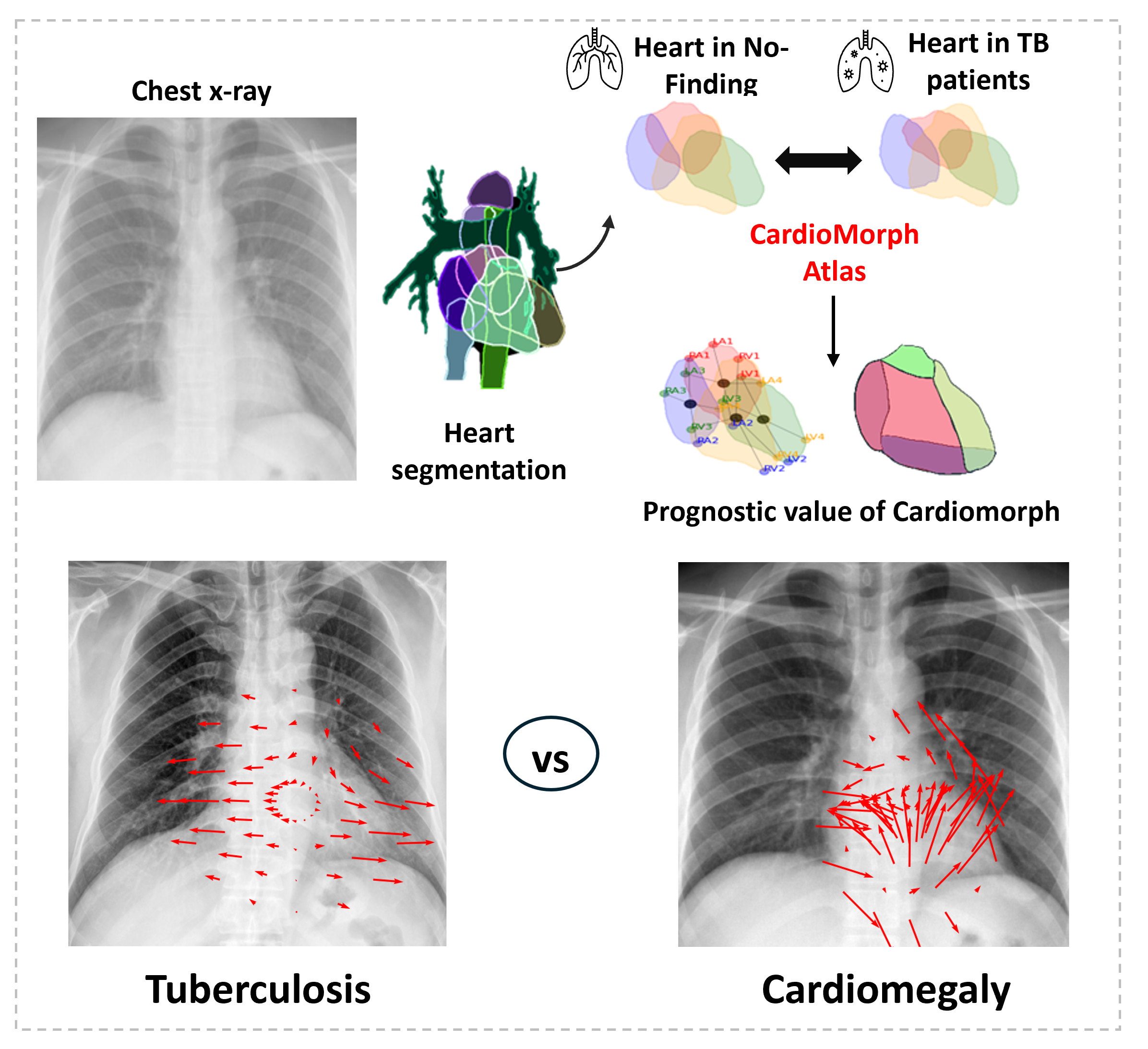
CardioMorph Atlas: A Statistical Approach to Evaluate Association between Pulmonary Tuberculosis and Cardiac Morphology from Chest X-rays Population-level statistical shape atlas linking tuberculosis to cardiac remodeling on chest X-raysAmritpal Singh, Sena Azamat, Gourav Modanwal, Pushkar Mutha, Pingfu Fu, Neel R. Gandhi, Rohan Dhamdhere, Mendel Lebowitz, Sadeer Al-Kindi, Anurag Aggarwal, Yingda L. Xie, Anant Madabhushi eBioMedicine (The Lancet Discovery Science) 2026;130:106385, 2026 Code / Paper / Project page / Background: Patients with tuberculosis (TB) face an elevated risk of certain cardiovascular diseases. The inflammatory response associated with TB is linked to cardiac complications, though the spatial impact on cardiac structure remains understudied. In this study, we assess population-level differences in cardiac morphology in TB using an atlas-based approach applied to routine chest X-rays. Methods: A total of 8,651 patients (2,626 with TB) were selected from four cohorts: Shenzhen, TBX11K, TB Portals, and MIMIC — covering seven countries. We developed a representative cardiac template, the CardioMorph Atlas, and compared population-level morphological differences between patients with TB and controls. Cardiac borders and areas were quantified into CardioMorph features and analysed for their association with all-cause mortality using Cox regression. Findings: CardioMorph Atlas revealed significant lateral elongation of the heart in patients with TB, in both left and right ventricles, with patterns distinct from those in pneumonia and cardiomegaly. Vector field analysis showed lateral extension of cardiac structures in patients with TB. Upper area (hazard ratio: 2.18, 95% CI: 1.10–4.30, p = 0.022) and right perimeter (HR = 2.07, 95% CI: 1.06–4.02) were significantly associated with increased all-cause mortality in patients with TB. Can a disease of the lungs leave a fingerprint on the heart’s outline, visible on the same 2D projection radiograph already being taken for TB screening? We built the CardioMorph Atlas by segmenting cardiac chambers and lung fields on 8,651 chest X-rays across four cohorts spanning seven countries, registering every heart to a median-area reference template (rigid + affine, DICE 0.841), converting chamber masks to signed distance functions, and running voxel-wise, permutation-corrected GLM comparisons (FSL |
|
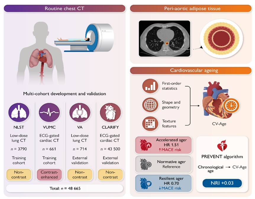
Peri-Aortic Fat to Assess Cardiovascular Aging Using an AI-Driven Radiomic Biomarker Peri-aortic adipose tissue radiomics on routine chest CT as a cardiovascular age-gap biomarkerMendel Lebowitz, Gourav Modanwal, Amritpal Singh, Rohan Dhamdhere, Saumya Diwan, Taofik Ahmed Suleiman, Hilmi Al-Shakhshir, Michael Gilkey, Scott Shofer, Santosh Kumar Sirasapalli, M Benjamin Shoemaker, Sadeer Al-Kindi, Sanjay Rajagopalan, Anant Madabhushi European Heart Journal 2026;ehag498, 2026 Paper / Background: Chronological age is central to cardiovascular disease (CVD) risk estimation but poorly reflects interindividual heterogeneity in cardiovascular aging. This study developed a peri-aortic adipose tissue (PAAT) radiomic cardiovascular age (CV-Age) biomarker from routine chest CT and tested whether the PAAT age gap (ΔAge = CV-Age − chronological age) stratifies major adverse cardiovascular events (MACE) and improves risk classification. Methods: Four chest CT cohorts with different protocols were used to train (n = 4,451) and externally validate (n = 44,214) a CV-Age model. Associations between ΔAge decile groups (resilient ≤10th; accelerated ≥90th) and incident 5-point MACE (myocardial infarction, stroke, revascularization, heart failure, and death) were assessed using survival models, with clinical utility evaluated by substituting CV-Age for chronological age in risk calculators. Findings: The PAAT-derived ΔAge identified patients at accelerated and resilient cardiovascular aging independent of chronological age, with accelerated agers showing significantly elevated 5-point MACE risk and resilient agers showing significantly lower risk, improving risk classification beyond standard clinical models. Chronological age is a blunt instrument for cardiovascular risk — two 60-year-olds can have wildly different vascular futures. We asked whether the fat wrapped around the aorta, already sitting in millions of chest CTs taken for unrelated reasons, encodes a better readout of cardiovascular age than the calendar does. We built a radiomic CV-Age model from peri-aortic adipose tissue (PAAT) segmented on chest CT, trained across 4,451 patients and externally validated on 44,214 across four cohorts with heterogeneous acquisition protocols. The target wasn’t age itself but the gap: ΔAge = CV-Age − chronological age, splitting the population into resilient agers (ΔAge in the bottom decile) and accelerated agers (top decile). Using landmark survival models for 5-point MACE (MI, stroke, revascularization, heart failure, death), accelerated PAAT-agers carried significantly elevated event risk and resilient agers significantly lower risk, holding after adjustment for standard clinical covariates — and substituting CV-Age for chronological age inside existing risk calculators improved classification. The result is a biomarker that piggybacks entirely on imaging already being acquired, turning an incidental fat depot into a personalized aging clock rather than adding a new acquisition, cohort, or protocol to the clinical pipeline. |
|
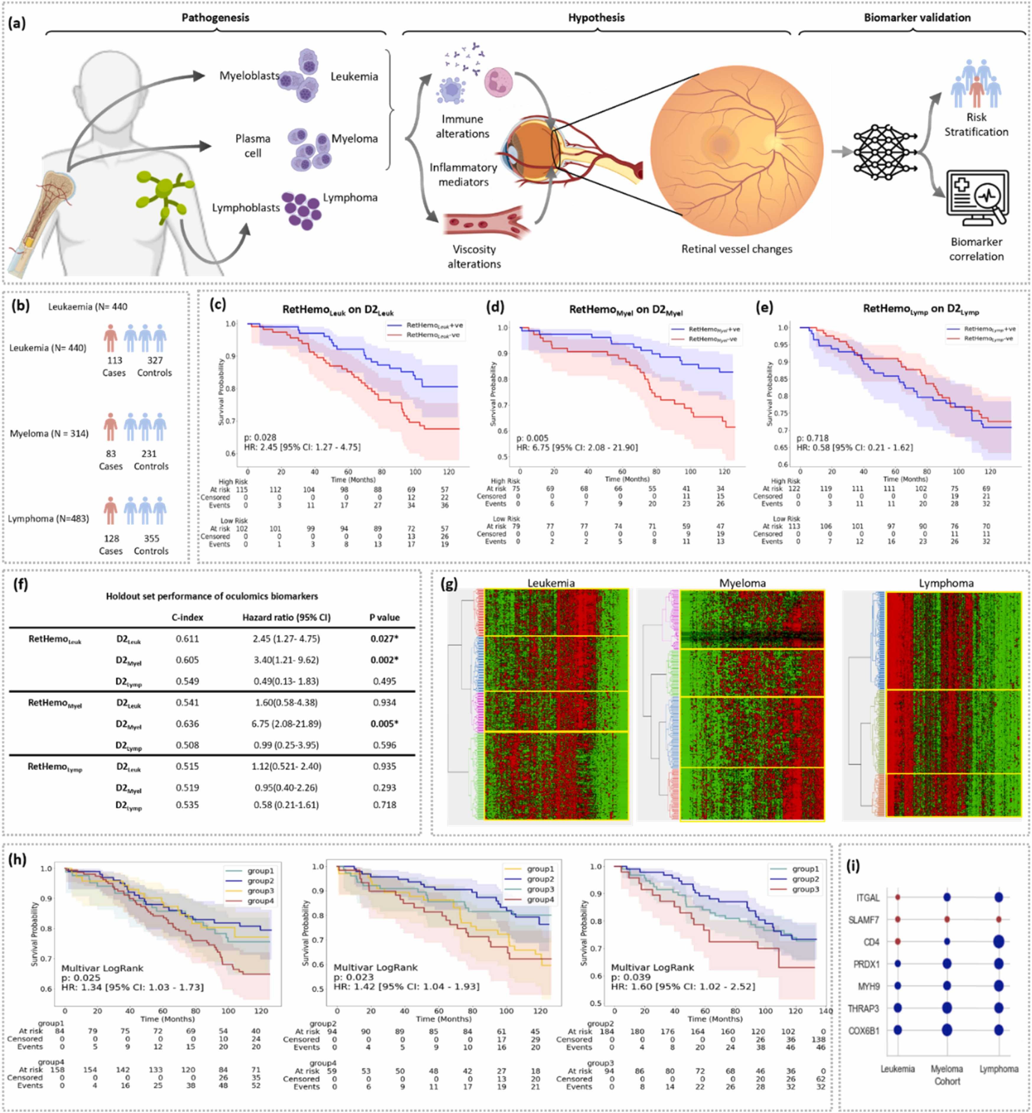
AI-informed retinal biomarkers predict 10-year risk of onset of multiple hematological malignancies Vasculature biomarker extraction for cancer detectionAmritpal Singh, Ajay K. Nooka, Gourav Modanwal, Nieraj Jain, Madhav V. Dhodapkar, Sruthi Arepalli, Sagar Lonial, Anant Madabhushi European Journal of Cancer, 2025 Paper / Project page / Background: Early detection of hematological malignancies improves long-term survival but remains a critical challenge due to heterogeneity in clinical presentation. Chronic inflammation is a key driver in hematologic cancers and is known to induce compensatory microvascular changes. High-resolution, non-invasive retinal imaging can allow the quantification of microvascular changes for the early detection of hematological malignancies. Methods and Results: RetHemo demonstrated significant predictive performance for leukemia (c-index = 0.611, HR = 2.45, 95 % CI: 1.27–4.75, p = 0.027), myeloma (c-index = 0.636, HR = 6.69, 95 % CI: 2.06–21.65, p = 0.006). Unsupervised hierarchical clustering based on retinal vasculature features identified distinct high-risk subgroups for leukemia (p = 0.013), myeloma (p < 0.001), and lymphoma (p = 0.034). Serum proteomics analysis revealed significantly elevated levels of inflammatory proteins, including ITGAL and SLAMF7, in high-risk patients. Comparison with clinical variables showed that RetHemo outperformed traditional clinical and hematologic parameters in stratifying at-risk individuals. Can hematologic malignancies really be “seen” in a structure as optically distant as the retina, or are we just overfitting noise across biology’s most vascular tissue? We force fundus images from 1,237 UK Biobank subjects into a vessel-centric representation—curvature, tortuosity, branching geometry—then compress it into a Cox latent risk axis that behaves less like a classifier and more like a time-to-event deformation field of microvasculature. Surprisingly, this latent space separates leukemia and myeloma trajectories years before diagnosis, but collapses for lymphoma, suggesting that systemic hematopoietic dysregulation imprints differently depending on marrow vs nodal dominance. Cross-testing exposes a brittle but structured transfer: leukemia signatures partially generalize into myeloma risk, hinting at shared inflammatory geometry, while myeloma-specific signals remain orthogonal, likely driven by paraprotein-induced rheology shifts. The model’s most unexpected behavior is not prediction accuracy, but that clustering alone recovers proteomic shifts in adhesion and immune signaling (ITGAL, SLAMF7), implying the retina is acting as a low-dimensional readout of circulating immune–vascular coupling rather than a disease-specific detector. |
|
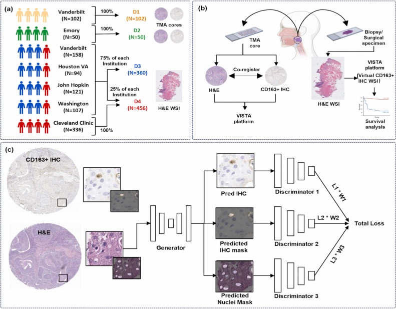
Artificial intelligence-based virtual staining platform for identifying tumor-associated macrophages from hematoxylin and eosin-stained images Image to Image translation: Virtual staining for Marcophage detectionArpit Aggarwal, Mayukhmala Jana, Amritpal Singh, Tanmoy Dam, Himanshu Maurya, Tilak Pathak, Sandra Orsulic, Kailin Yang, Deborah Chute, Justin A Bishop, Farhoud Faraji, Wade M Thorstad, Shlomo Koyfman, Scott Steward, Qiuying Shi, Vlad Sandulache, Nabil F Saba, James S Lewis Jr, Germán Corredor, Anant Madabhushi European Journal of Cancer, 2025 Paper / Background: Virtual staining is an artificial intelligence-based approach that transforms pathology images between stain types, such as hematoxylin and eosin (H&E) to immunohistochemistry (IHC), providing a tissue-preserving and efficient alternative to traditional IHC staining. However, existing methods for translating H&E to virtual IHC often fail to generate images of sufficient quality for accurately delineating cell nuclei and IHC+ regions. To address these limitations, we introduce VISTA, an artificial intelligence-based virtual staining platform designed to translate H&E into virtual IHC.Methods and Results: We applied VISTA to identify M2-subtype tumor-associated macrophages (M2-TAMs) in H&E images from 968 patients with HPV+ oropharyngeal squamous cell carcinoma across six institutional cohorts. Co-registered H&E and CD163 + IHC tissue microarrays were used to train (D1, N = 102) and test (D2, N = 50) the VISTA platform. M2-TAM density, defined as the ratio of M2-TAMs to total nuclei. High M2-TAM density was associated with worse overall survival in D4 (p = 0.0152, Hazard Ratio=1.63 [1.1–2.42]). VISTA outperformed existing methods, generating higher-quality virtual CD163 + IHC images in D2, with a Structural Similarity Index of 0.72, a Peak Signal-to-Noise Ratio of 21.5, and a Fréchet Inception Distance of 41.4. Additionally, VISTA demonstrated superior performance in segmenting M2-TAMs in D2 (Dice=0.74). Pathologists see immune cells everywhere in H&E slides. The problem is that identifying specific phenotypes like CD163+ tumor-associated macrophages still usually requires an additional IHC stain, extra tissue sections, added cost, and a slower workflow. So we asked a simple question: Can a model learn the mapping from morphology in H&E directly to immunophenotype-specific staining patterns? VISTA — a multi-task virtual staining framework that translates H&E into virtual CD163 IHC while simultaneously generating: macrophage-positive segmentation masks along with high-fidelity synthetic IHC. The system combines conditional GANs with a shared multi-objective optimization framework. Instead of treating image translation and segmentation independently, VISTA jointly optimizes all three tasks together using ResNet-based generators for stain translation, U-Net generators for segmentation, PatchGAN discriminators for high-frequency structural realism along with hybrid adversarial + Huber + segmentation objectives. The pipeline was trained using co-registered H&E/CD163 tissue microarrays and then deployed on large-scale whole slide cohorts across multiple institutions. Compared against Pix2Pix, CycleGAN, CUT, FastCUT, PyramidPix2Pix, and PSPStain, VISTA achieved stronger reconstruction fidelity and better macrophage segmentation performance. Even more interesting: the derived M2-TAM density biomarker from virtual staining remained prognostic for overall survival in HPV+ OPSCC. |
|
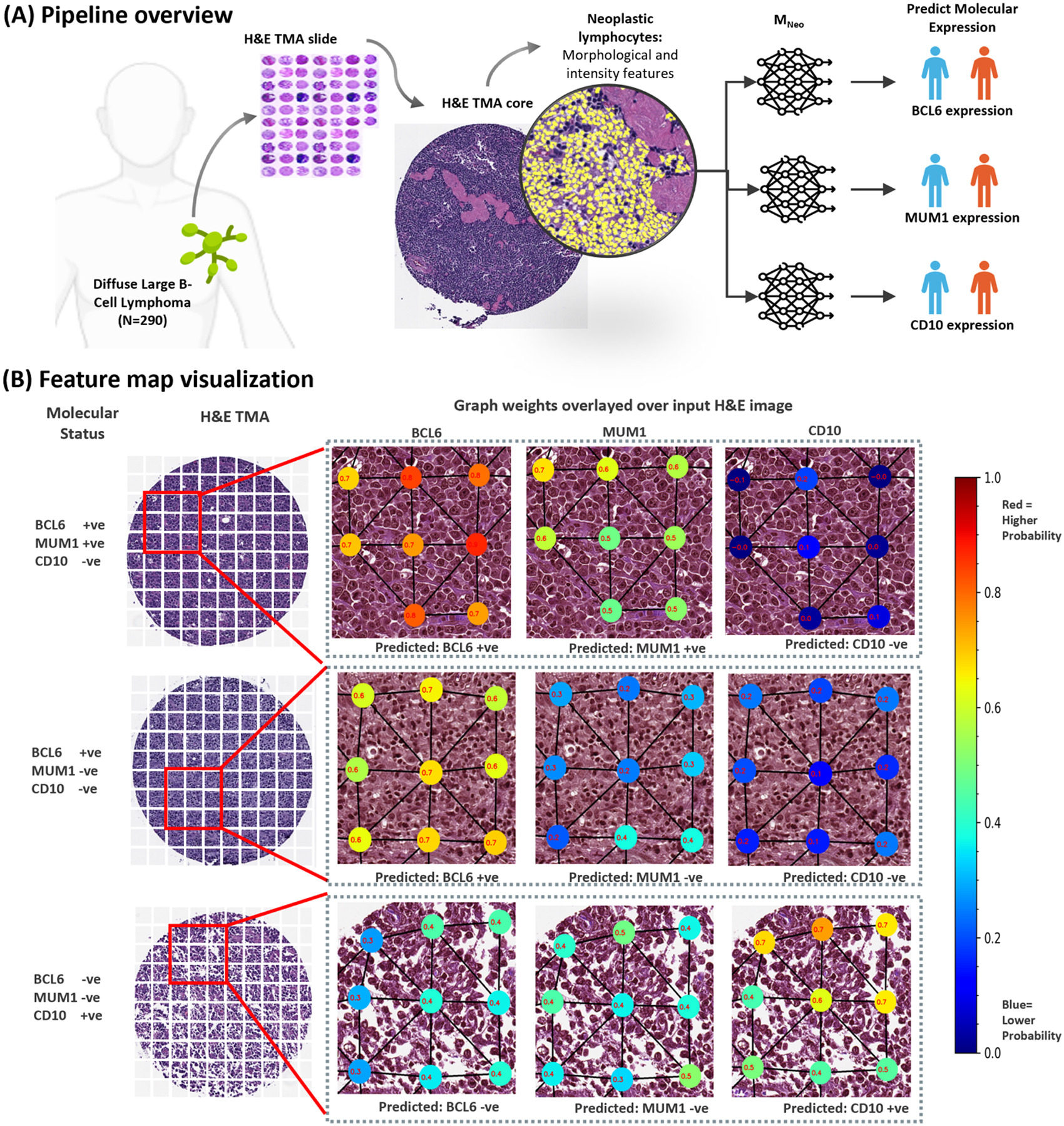
Spatial Arrangement of Neoplastic Lymphocytes Predicts Molecular Subtypes in Diffuse Large B-cell Lymphoma Graph Neural Networks for Spatial Arrangement of Neoplastic Lymphocytes in DLBCLAmritpal Singh, Tilak Pathak, Germán Corredor, Anant Madabhushi Oral talk, USCAP (USA), 2025 Paper / Diffuse large B-cell lymphoma (DLBCL) is the most common non-Hodgkin lymphoma and can be fatal if untreated. Accurate cell-of-origin (COO) classification is critical for prognosis and treatment, but standard immunohistochemistry (IHC) for BCL6, MUM1, and CD10 is costly, time-consuming, and not widely accessible. Alternative approaches using routine histopathology could improve scalability and access. We analyzed digitized H&E tissue microarray images from DLBCL patients. Images were tiled, neoplastic lymphocytes segmented with Hover-Net, and nuclear morphology and intensity features extracted. A graph neural network, MNeo, was trained to predict BCL6, MUM1, and CD10 expression, compared to a baseline foundation model. MNeo reliably predicted molecular subtypes from H&E images, outperforming the baseline. Feature maps highlighted regions most informative for each marker, showing that nuclear morphology patterns carry meaningful molecular information. This study demonstrates that graph-based computational pathology can serve as a cost-effective, accessible alternative to IHC, enhancing diagnostic efficiency and enabling broader application of precision medicine in DLBCL. Under the microscope, DLBCL can look deceptively similar across patients — but the spatial organization of lymphocytes tells a much richer molecular story. In this work, we modeled lymphoma tissue as a graph. Using Hover-Net nuclei segmentation, handcrafted nuclear morphology/intensity features, and Delaunay-based graph construction, we trained GNNs to predict molecular subtype markers directly from routine H&E slides. The interesting part was that the model was not just learning individual cells — it was learning neighborhood structure, tissue topology, and spatial cellular interactions linked to BCL6, MUM1, and CD10 expression. Our graph-based framework, MNeo, outperformed baseline foundation models and showed that spatial lymphocyte architecture itself encodes latent molecular phenotype information. Presented at the USCAP 2025. |
|
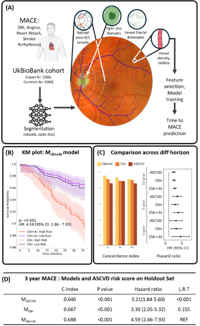
Explainable AI Better Predicts 3-Year MACE Risk Compared to Clinical and ASCVD Models in the UK Biobank CohortAmritpal Singh, Rohan Dhamdhere, Gourav Modanwal, Sudeshna Sil Kar, Sadeer Al-Kindi, Anant Madabhushi American Heart Association (AHA), 2024 Paper / Cardiovascular risk prediction could be improved by analyzing retinal microvascular architecture, as standard risk calculators may miss early vascular changes. This study aimed to assess whether AI-derived retinal vessel features could predict 3-year MACE and improve upon traditional risk models. We analyzed baseline fundus images from 2,120 UK Biobank participants without prior CVD, extracting vessel features such as angle, tortuosity, curvature, and caliber. Cox models incorporating demographics, clinical factors, and AI-derived retinal features were trained and validated to predict MACE risk. AI-derived retinal features significantly improved prediction of 3-year MACE compared to clinical factors alone and the ASCVD risk calculator. Integrated models highlighted novel vascular patterns captured from fundus images that correlated with event risk. This approach demonstrated the greatest predictive power in the short term, suggesting retinal imaging can provide complementary information to existing risk assessments. The findings support AI-based retinal biomarkers as a non-invasive, accessible tool for enhanced cardiovascular risk stratification, with potential for prospective multi-center validation. |
|
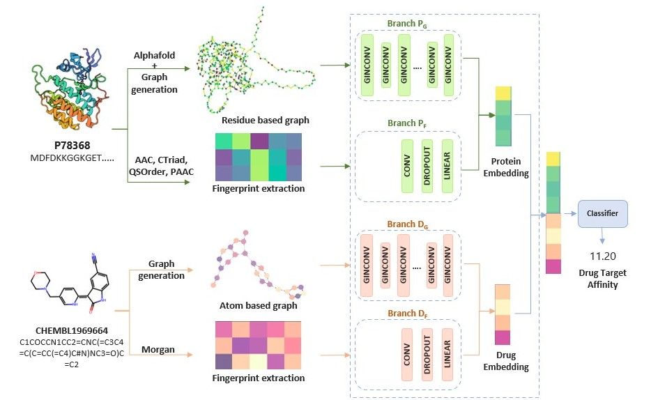
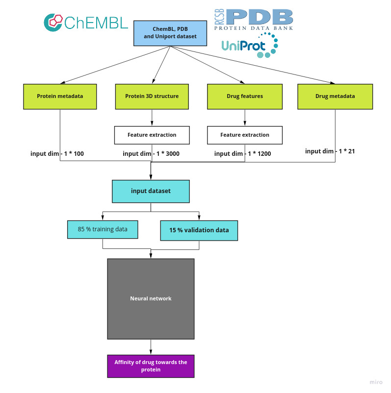
GraphPrint: Combining Traditional Fingerprint with Graph Neural Networks For Drug Target Prediction GraphPrint: Combining Traditional Fingerprint with Graph Neural Networks For Drug Target PredictionAmritpal Singh NEURIPS, 2023 Code / Paper / Project page / Accurate drug target affinity prediction can improve drug candidate selection, accelerate the drug discovery process, and reduce drug production costs. Previous work focused on traditional fingerprints or used features extracted based on the amino acid sequence in the protein, ignoring its 3D structure which affects its binding affinity.In this work, we propose GraphPrint: a framework for incorporating 3D protein structure features for drug target affinity prediction. We generate graph representations for protein 3D structures using amino acid residue location coordinates and combine them with drug graph representation and traditional features to jointly learn drug target affinity. Our model achieves a mean square error of 0.1378 and a concordance index of 0.8929 on the KIBA dataset and improves over using traditional protein features alone. Our ablation study shows that the 3D protein structure-based features provide information complementary to traditional features. pdf paper Can a protein’s function really be learned from its sequence if the physics is encoded in its 3D folding geometry, not its letters? We construct GraphPrint by collapsing AlphaFold-derived protein structures into residue-level graphs, where each node is not just an amino acid but a geometric event in 3D space carrying latent physicochemical constraints. The model then fuses this structural manifold with ligand graphs, but the interesting behavior is not fusion—it is redundancy breakdown, where sequence fingerprints and structure embeddings stop agreeing and begin encoding orthogonal binding priors. On KIBA, the system does not just improve correlation; it exposes a sharp failure mode where sequence-only baselines systematically mis-rank large, aromatic, high-bond-count ligands, suggesting that binding affinity errors scale with molecular complexity in a way graph convolutions alone cannot linearize. The key signal is that adding 3D geometry does not “add information” in a simple sense—it reshapes the protein representation space so that docking behavior becomes a graph alignment problem rather than a language modeling problem. |

|
Class-Incremental Continual Learning for General Purpose Healthcare Models Class-Incremental Continual Learning for General Purpose Healthcare ModelsAmritpal Singh, Mustafa Burak Gurbuz, Shiva Souhith Gantha, Prahlad Jasti NEURIPS, 2023 Paper / Project page / Healthcare clinics regularly encounter dynamic data that changes due to variations in patient populations, treatment policies, medical devices, and emerging disease patterns. Deep learning models can suffer from catastrophic forgetting when fine-tuned in such scenarios, causing poor performance on previously learned tasks. Continual learning allows learning on new tasks without performance drop on previous tasks. In this work, we investigate the performance of continual learning models on four different medical imaging scenarios involving ten classification datasets from diverse modalities, clinical specialties, and hospitals. We implement various continual learning approaches and evaluate their performance in these scenarios. Our results demonstrate that a single model can sequentially learn new tasks from different specialties and achieve comparable performance to naive methods. These findings indicate the feasibility of recycling or sharing models across the same or different medical specialties, offering another step towards the development of general-purpose medical imaging AI that can be shared across institutions. Can a single model remember what it learned from dermatology, pathology, and radiology without quietly overwriting its own past? We simulate sequential exposure to ten heterogeneous medical imaging tasks across hospitals, modalities, and specialties, where each new dataset acts like a distributional shock that destabilizes previously learned feature hierarchies. The interesting failure mode is not accuracy on new tasks, but representational drift: shared CNN backbones either fragment into task-specific subspaces or collapse into overly conservative features that forget fine-grained medical cues. Replay-based continual learning (especially MAS+r and DER++) stabilizes this drift, but in a non-uniform way-some tasks act as anchors while others behave like destructive interference patterns in the latent space. What emerges is not a “general-purpose medical model,” but a constrained plasticity system where forgetting is not eliminated, only redistributed across tasks depending on how compatible their visual statistics are. |

|
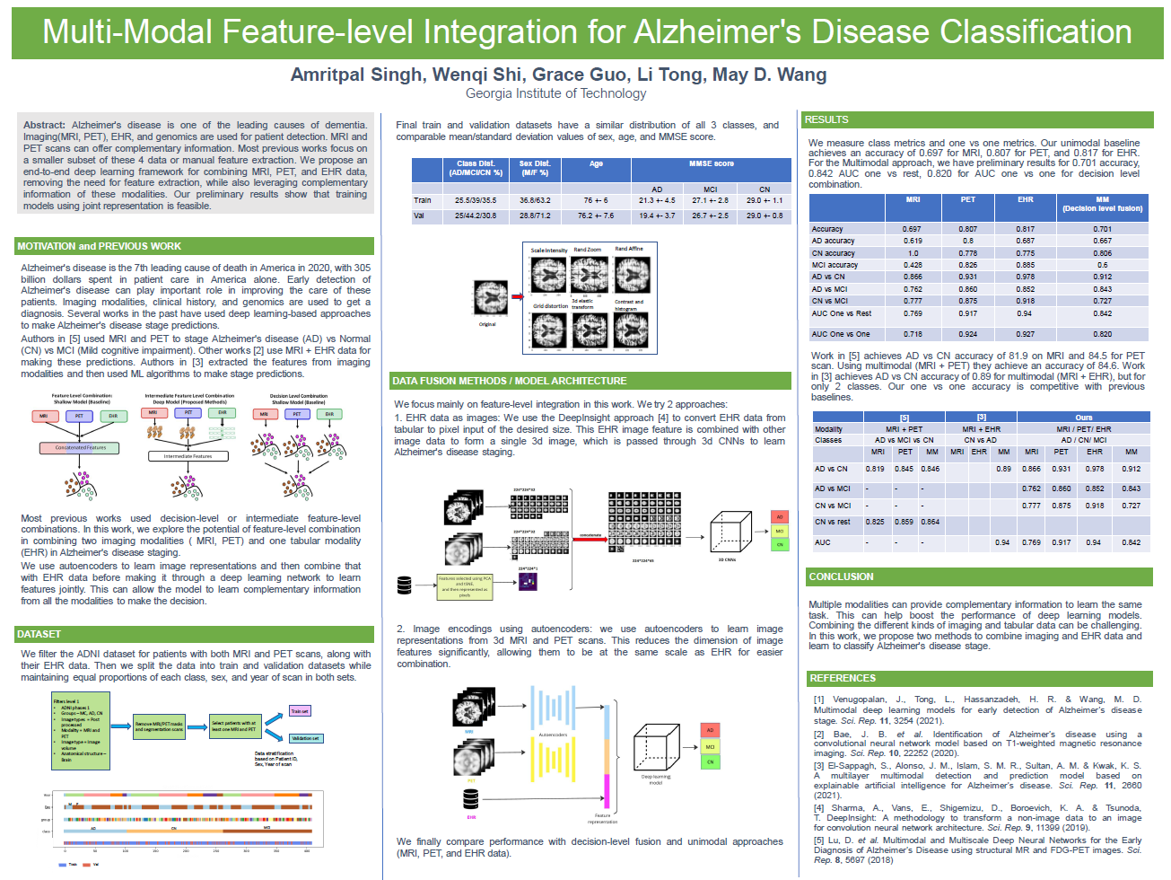
Multi-Modal Deep Feature Integration for Alzheimer’s Disease Staging Multi-Modal Deep Feature Integration for Alzheimer’s Disease StagingAmritpal Singh, Wenqi Shi, May D. Wang IEEE BIBM conference, 2023 Paper / Project page / Alzheimer’s disease (AD) is one of the leading causes of dementia and 7th leading cause of death in the United States. The provisional diagnosis of AD relies on comprehensive examinations, including medical history, neurological and psychiatric examinations, cognitive assessments, and neuroimaging studies. Integrating diverse sets of clinical data, including electronic health records (EHRs), medical imaging, and genomic data, enables a holistic view of AD staging analysis. In this study, we propose an end-to-end deep learning architecture to jointly learn from magnetic resonance imaging (MRI), positron emission tomography (PET), EHRs, and genomics data to classify patients into AD, mild cognitive disorders, and controls. We conduct extensive experiments to explore different feature-level and intermediate-level fusion methods. Our findings suggest intermediate multiplicative fusion achieves the best stage prediction performance on the external validation dataset. Compared with unimodal baselines, we can observe that integrative approaches that leverage all four modalities demonstrate superior performance to baselines reliant solely on one or two modalities. In an age-wise comparison, we observe a unique pattern that all fusion methods exhibited superior performance in the earlier age brackets (50-70 years), with performance diminishing as the age group advanced (70-90 years). The proposed integration framework has the potential to augment our understanding of disease diagnosis and progression by leveraging complementary information from multimodal patient data. What happens when you force MRI scans, PET metabolism maps, EHR timelines, and genomic signals into the same latent space and ask them to agree on whether a brain is aging normally or collapsing toward Alzheimer’s? This work builds a multimodal deep learning framework that treats Alzheimer’s staging as a representation alignment problem across radically different biomedical modalities. Surprisingly, simple multiplicative fusion consistently beat larger concatenative models, suggesting that cross-modal interaction mattered more than parameter count. The study also uncovered a strange age effect: models became highly predictive in earlier disease windows, then progressively failed in older cohorts, hinting at hidden subtypes of neurodegenerative aging. We even transformed structured clinical/genomic variables into image-like spatial embeddings to co-train with 3D neuroimaging pipelines. The result is less of a medical classifier and more of a small-scale experiment in multimodal reasoning over noisy human biology. |
|
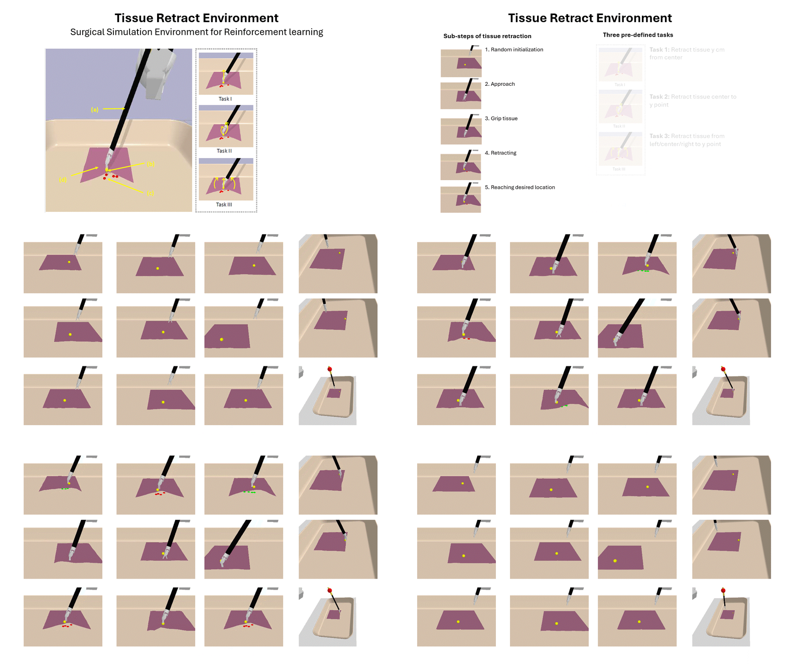
Autonomous Soft Tissue Retraction Using Demonstration-Guided Reinforcement Learning Autonomous Soft Tissue Retraction Using Demonstration-Guided Reinforcement Learning mAmritpal Singh, Wenqi Shi, May D. Wang MICCAI conference, 2023 Code / Paper / Project page / In the context of surgery, robots can provide substantial assistance by performing small, repetitive tasks such as suturing, needle exchange, and tissue retraction, thereby enabling surgeons to concentrate on more complex aspects of the procedure. However, existing surgical task learning mainly pertains to rigid body interactions, whereas the advancement towards more sophisticated surgical robots necessitates the manipulation of soft bodies. Previous work focused on tissue phantoms for soft tissue task learning, which can be expensive and can be an entry barrier to research. Simulation environments present a safe and efficient way to learn surgical tasks before their application to actual tissue. In this study, we create a Robot Operating System (ROS)-compatible physics simulation environment with support for both rigid and soft body interactions within surgical tasks. Furthermore, we investigate the soft tissue interactions facilitated by the patient-side manipulator of the DaVinci surgical robot. Leveraging the pybullet physics engine, we simulate kinematics and establish anchor points to guide the robotic arm when manipulating soft tissue. Using demonstration-guided reinforcement learning (RL) algorithms, we investigate their performance in comparison to traditional reinforcement learning algorithms. Our in silico trials demonstrate a proof-of-concept for autonomous surgical soft tissue retraction. The results corroborate the feasibility of learning soft body manipulation through the application of reinforcement learning agents. This work lays the foundation for future research into the development and refinement of surgical robots capable of managing both rigid and soft tissue interactions. What if surgical robots could learn soft tissue manipulation before ever touching a patient? This project drops a DaVinci surgical robot into a physics simulation where it has to figure out one of the hardest parts of surgery: handling deformable soft tissue. Not rigid tools. Not fixed objects. Realistic tissue that stretches, slips, recoils, and fights back. We built a ROS-compatible soft-body simulation environment using PyBullet and trained reinforcement learning agents to autonomously perform tissue retraction tasks. The system combines demonstration-guided RL with surgical robotics, allowing agents to learn from expert-style guidance while still exploring on their own. The interesting part: the robot gradually learns behaviors almost like a trainee surgeon — first approaching tissue, then grasping anchor points, then learning how to pull without losing grip or damaging the tissue. We also compared pure RL vs demonstration-guided RL across multiple soft-tissue manipulation tasks and studied how the number of demonstrations changes performance. This work was accepted at MICCAI 2023 AE-CAI Workshop. |
|
Multi-Modality Deep Learning Methods to Learn Alzheimer’s Disease ClassificationAmritpal Singh, Wenqi Shi, May D. Wang Georgia Institute of Technology, 2023 |
|
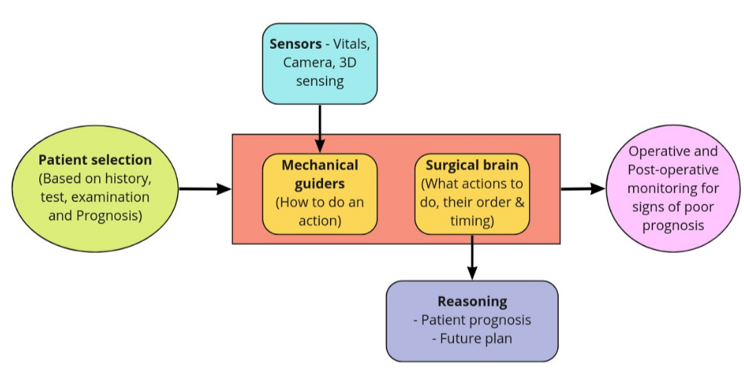
Roadmap to Autonomous Surgery - A Framework to Surgical AutonomyAmritpal Singh arxiv, 2022 Paper / Robotic surgery has increased the domain of surgeries possible. Several examples of partial surgical automation have been seen in the past decade. We break down the path of automation tasks into features required and provide a checklist that can help reach higher levels of surgical automation. Finally, we discuss the current challenges and advances required to make this happen. |
|
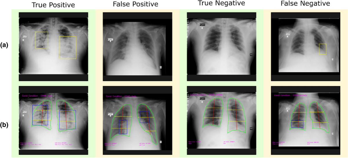
Validation of expert system enhanced deep learning algorithm for automated screening for COVID-Pneumonia on chest X-raysPrashant Sadashiv Gidde, Shyam Sunder Prasad, Ajay Pratap Singh, Nitin Bhatheja, Satyartha Prakash, Prateek Singh, Aakash Saboo, Rohit Takhar, Salil Gupta, Sumeet Saurav, Raghunandanan M. V., Amritpal Singh, Viren Sardana, Harsh Mahajan, Arjun Kalyanpur, Atanendu Shekhar Mandal, Vidur Mahajan, Anurag Agrawal, Anjali Agrawal, Vasantha Kumar Venugopal, Sanjay Singh & Debasis Dash Nature, Scientific Reports, 2021 Paper / SARS-CoV2 pandemic exposed the limitations of artificial intelligence based medical imaging systems. Earlier in the pandemic, the absence of sufficient training data prevented effective deep learning (DL) solutions for the diagnosis of COVID-19 based on X-Ray data. Here, addressing the lacunae in existing literature and algorithms with the paucity of initial training data; we describe CovBaseAI, an explainable tool using an ensemble of three DL models and an expert decision system (EDS) for COVID-Pneumonia diagnosis, trained entirely on pre-COVID-19 datasets. The performance and explainability of CovBaseAI was primarily validated on two independent datasets. Firstly, 1401 randomly selected CxR from an Indian quarantine center to assess effectiveness in excluding radiological COVID-Pneumonia requiring higher care. Second, curated dataset; 434 RT-PCR positive cases and 471 non-COVID/Normal historical scans, to assess performance in advanced medical settings. CovBaseAI had an accuracy of 87% with a negative predictive value of 98% in the quarantine-center data. However, sensitivity was 0.66–0.90 taking RT-PCR/radiologist opinion as ground truth. This work provides new insights on the usage of EDS with DL methods and the ability of algorithms to confidently predict COVID-Pneumonia while reinforcing the established learning; that benchmarking based on RT-PCR may not serve as reliable ground truth in radiological diagnosis. Such tools can pave the path for multi-modal high throughput detection of COVID-Pneumonia in screening and referral. |
|
Personalized Brain State Targeting via Reinforcement LearningAbhishek Naik, Amritpal Singh, Koushani Biswas, Harini Sudha, Matthew Schlegel, Kyle E. Mathewson Neuromatch academy, 2020 website / We propose a novel use of reinforcement learning as an active closed-loop assistive system that learns in real time to lead any brain state to a given goal state. Previous open- and closed-loop systems to manipulate brain states are generally passive in the sense that they are trained offline from data collected from a population but are not tailored or adapted for any individuals. Offline adaptation per individual if at all is very slow. Adaptation, and the speed of adaptation, is critical in most applications where manipulation of brain states is performed because poor initial performance and long training periods are barriers to BCI adaptation and success. Reinforcement learning is a sequential decision-making paradigm in which the system learns to map situations to optimal actions via trial-and-error interactions with the world to maximize a reward signal. Crucially, this reward signal is a form of evaluative feedback, for instance, proportional to how far the current state is from the goal state. This is in contrast to instructive feedback in the supervised learning paradigm, where the correct action is assumed known. We propose modeling brain state manipulation as a sequential decision-making problem, wherein a system takes real-time EEG data as input and uses audio-visual cues to start from any brain state and reach a physiologically-objective goal state such as a particular oscillation frequency or a deep-sleep state. We show a proof of concept example using a consumer-based EEG device. We believe such an active closed-loop system would have a large impact in assistive applications ranging from helping critically-ill patients fall asleep to helping everyday stressed-out individuals relax. |
|
In-Silico Repositioning of Drugs for Neurofibromatosis 2 Vestibular Schwannoma using Machine LearningMIT Hack 4 Rare Disease Hackathon, 2020 website / Neurofibromatosis type 2 is an autosomal-dominant multiple neoplasia syndrome. It is highly debilitating with the frequency of one in 25,000 live births and nearly 100% penetrance by 60 years of age (1). NF2 represents a difficult management problem with most patients facing substantial morbidity and reduced life expectancy. The hallmark of NF2 is the appearance of bilateral vestibular schwannomas, benign tumors on both sides of the vestibular nerve. People with NF2 may also develop schwannomas in other parts of the body, or may develop other types of benign brain or spinal tumors (2) .At this time, possible treatments available for Neurofibromatosis 2-associated tumors include surgery, chemotherapy, and radiation therapy (3). Presently available off-label drugs are not fully effective. It has also been observed that monotherapy is not effective with lesser efficacy, higher resistance and toxicity. The complex interlinked pathways in the pathogenesis NF2 suggest multi-drug therapy may provide an ideal therapeutic effect (4). We therefore would like to work on developing a Machine Learning model to identify suitable drug combination that could lead to better efficacy and lesser chance of resistance. |
ProjectsThese include coursework and side projects. |

|
Optimize Task Allocation via Redundancy in Multi-Agent Systemsprojects 2022-12-10 Task allocation is an important problem to be solved. Several real life scenarios require efficient task alloacation, like:
|

|
Path planning to control robotic arm for suturingprojects 2022-12-06 Project for CS-6739 Medical robotics course, MSCS Gatech, USA Team members: Amritpal Singh, Oluwatofunmi M Sodimu Group 5 (G) , BMED 6739 Medical robotics Advisor - Prof. Yue Chen |
|
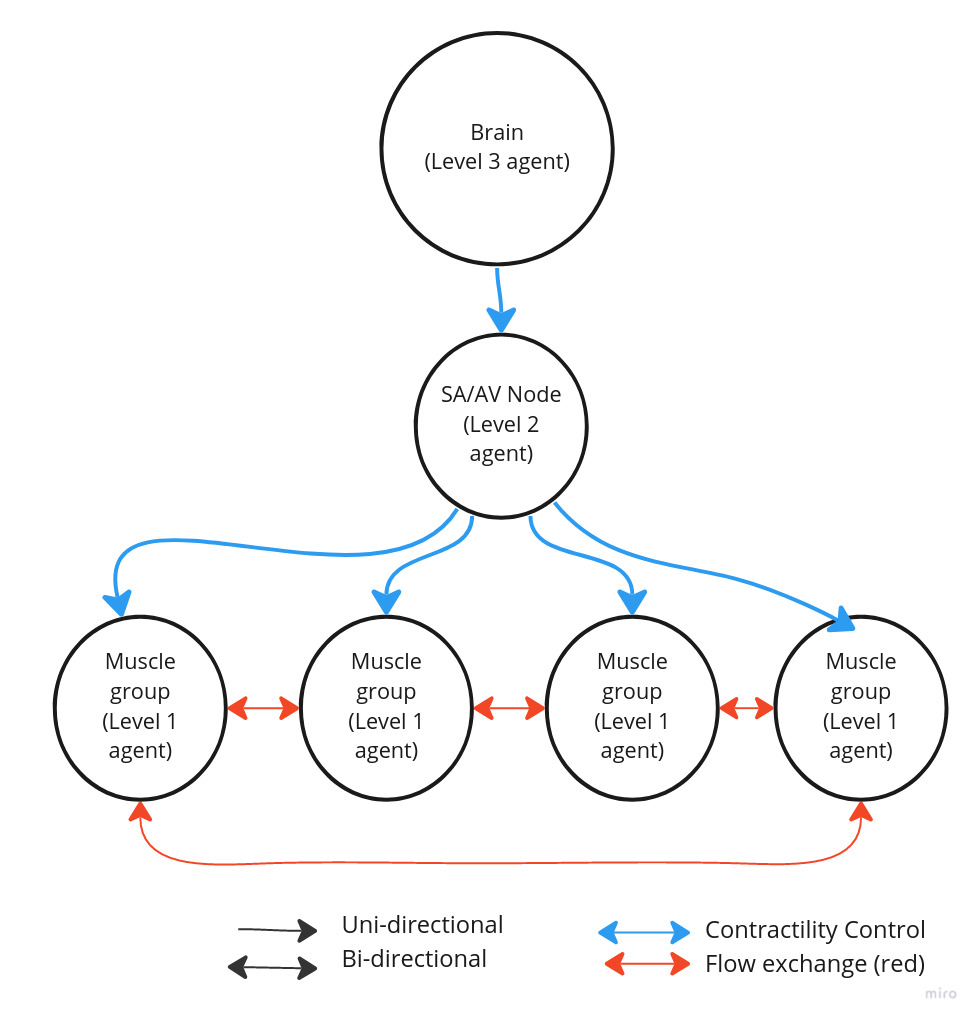
Efficient blood pumping in Bionic heart via distributed control as Multi-agent systemprojects 2022-10-25 Decentralised control of heart muscles dynamics to allow efficient coordination for pumping blood. Programmed directed acyclic graph based reinforcement learning environment with vertices representing agents, and edges equal to blood flow. Two sub-problems to solve: 1. Calculate blood flow using mathematics of fluid dynamics and then using Ford-Fulkerson max flow algorithm to derive find max blood flow in graph at any time. 2. Reinforcement learning algo to solve for optimal coordination policy |
|
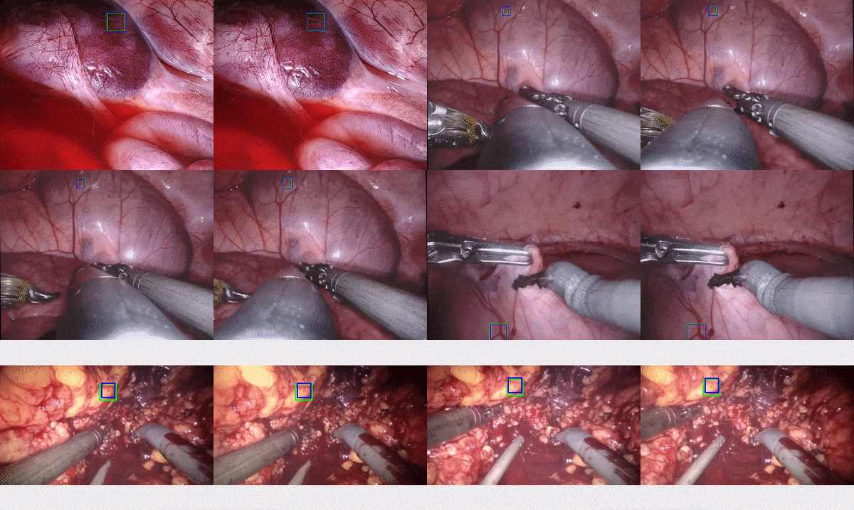
Surgical Tracking in endoscopic videosprojects 2022-09-15 Visual tracking involves following a bounding box throughout a video sequence. This is a crucial task in Computer-Assisted Interventions (CAI), with a range of applications including soft tissue deformation estimation, lesion tracking, augmented reality and robotic visual servoing. Medical applications require accurate trackers that are robust in challenging conditions prevalent in surgery. Hence prior to being utilized in real-world practice, tissue trackers need to be evaluated in large and diverse datasets that capture multiple challenging conditions. To address this, we propose the SurgT challenge, a new first-of-a-kind collection of tools and datasets for training and benchmarking tissue trackers in surgery. |
|
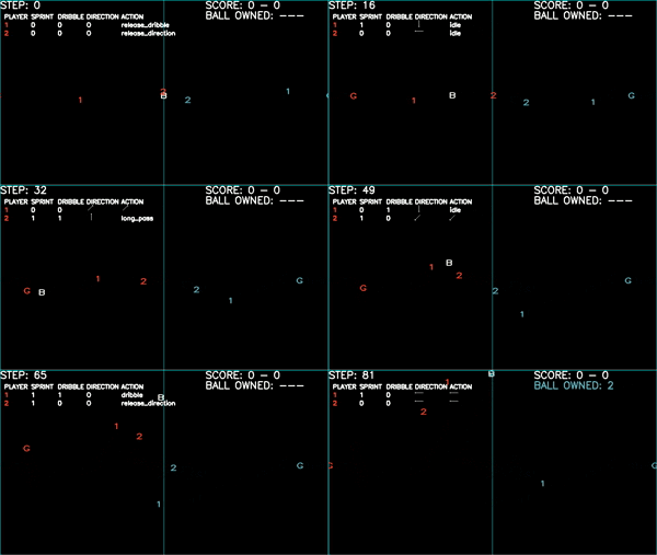
Football Qmix Multiagent agent efficient team coordination in Football using QMIX reinforcement learning algorithmprojects 2022-04-30 Code / Project for CS-7641, MS CS Gatech, USA. Challenges of Multi-agent reinforcement learning On top of the exploration-exploitation dilemma, Multi-agent RL faces another dilemma called the Predictability exploitation dilemma. Maximizing performance requires collecting rewards. As in Dec-POMDP agents cannot explicitly communicate, coordination requires predictability. At times, this predictability can also require ignoring private information. The dilemma is to choose between the benefit of exploiting private observation vs the cost of predictability. |
|
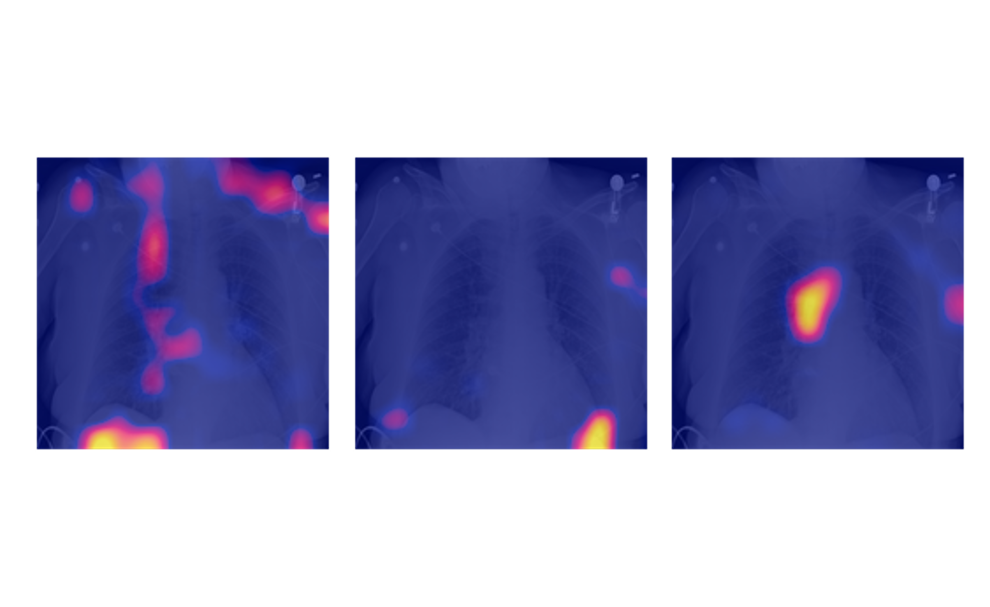
RANZCR CLiP - Catheter and Line Position Challengeprojects 2021-03-16 website / Classify the presence and correct placement of tubes on chest x-rays to save lives. Evaluation metric: Modified version of the Laplace Log Likelihood.
|

|
Vitals: Android app to track patient vitalsprojects 2021-01-01 website / Code / Demo version of app to track your vitals - Temp, BP, SP02, BP.
Track vitals over time, See graph representations.
|
|
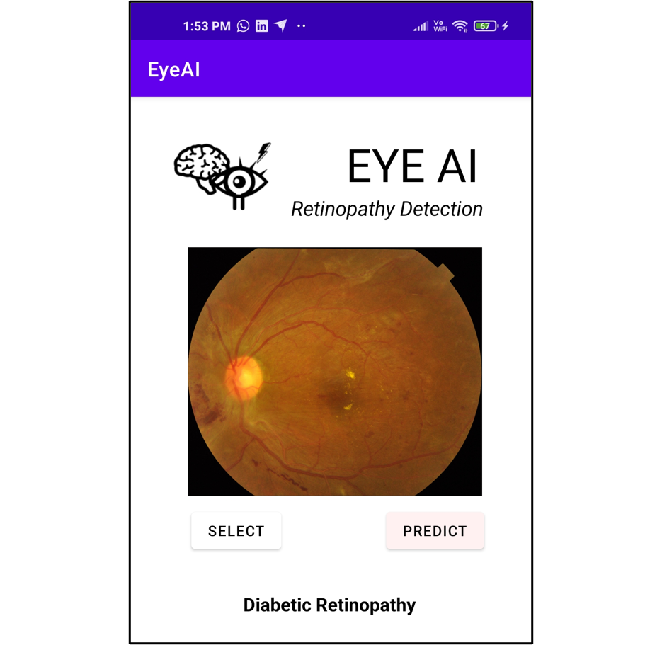
EyeAI - Android appprojects 2021-01-01 website / Code / Prototype Deep leanring based android app for eye disease on edge devices Training, deploying deep learning models for ophthalmology diagnosis on android app |
|
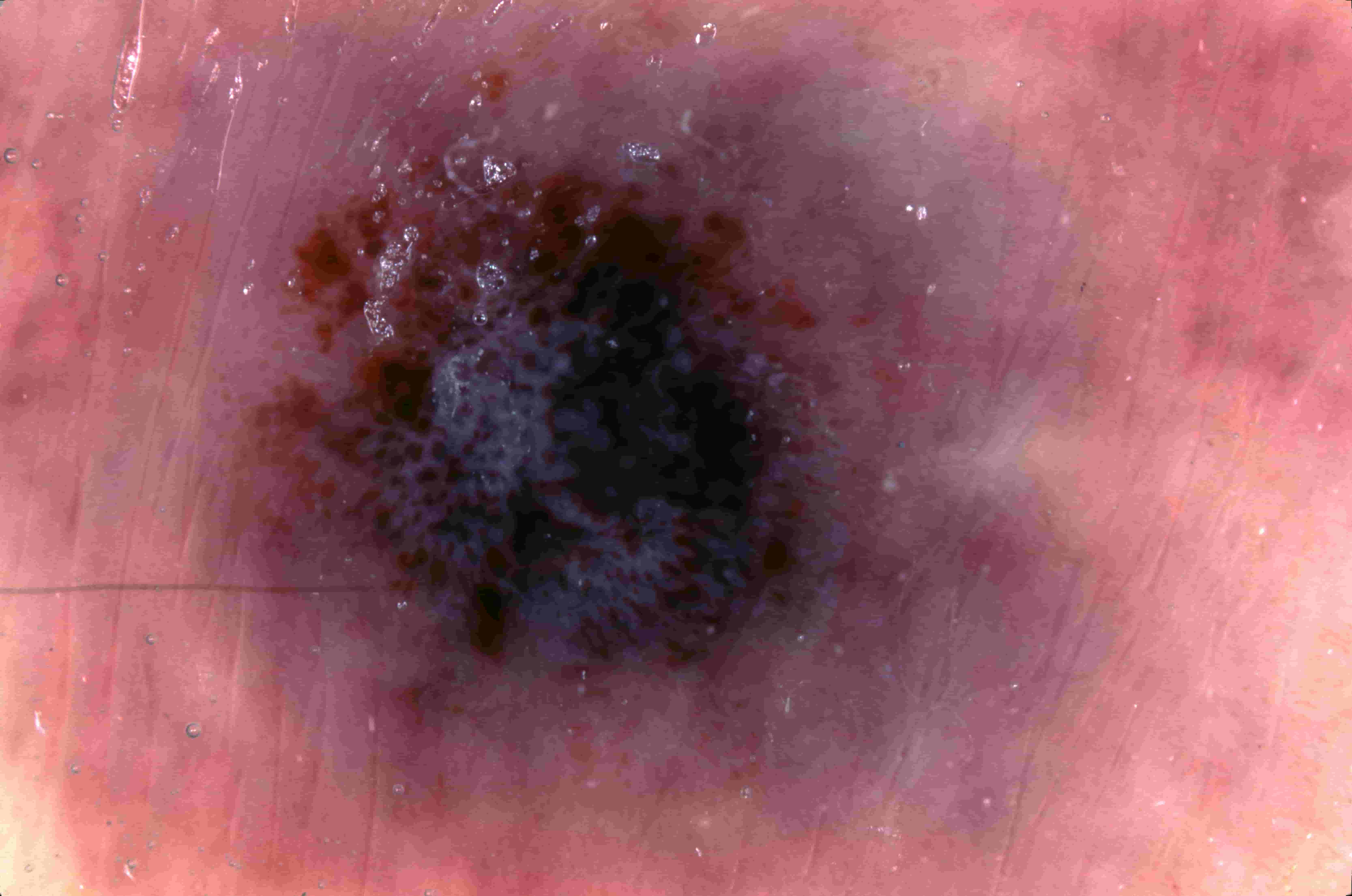
DermaAI - Deep learning based Skin lesions Classificationprojects 2021-01-01 Code / CNNs based classification of skin lesions Aim: Deploying deep learning models for Derma diagnosis |

Experience
|